Figure 1. [The normal human retina fundus]. - Webvision - NCBI
Por um escritor misterioso
Descrição
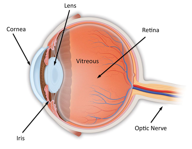
The normal human retina fundus photo shows the optic nerve (right), blood vessels and the position of the fovea (center).
![Figure 1. [The normal human retina fundus]. - Webvision - NCBI](https://pub.mdpi-res.com/symmetry/symmetry-15-01631/article_deploy/html/images/symmetry-15-01631-g002.png?1692867486)
Symmetry, Free Full-Text
![Figure 1. [The normal human retina fundus]. - Webvision - NCBI](https://www.researchgate.net/publication/333702798/figure/fig1/AS:771957922480128@1561060519865/The-human-retina-with-different-stages-of-NPDR-a-normal-retina-and-its-main-components.png)
The human retina with different stages of NPDR: a normal retina and its
![Figure 1. [The normal human retina fundus]. - Webvision - NCBI](https://journals.physiology.org/cms/10.1152/physrev.00035.2019/asset/images/medium/z9j004202952r001.png)
Emerging Approaches for Restoration of Hearing and Vision
![Figure 1. [The normal human retina fundus]. - Webvision - NCBI](https://www.cell.com/cms/attachment/75170c2e-8c9f-41dc-bb88-3db83ce1f63c/gr3_lrg.jpg)
Suprachoroidal and Subretinal Injections of AAV Using Transscleral Microneedles for Retinal Gene Delivery in Nonhuman Primates: Molecular Therapy - Methods & Clinical Development
![Figure 1. [The normal human retina fundus]. - Webvision - NCBI](https://eophtha.com/images/uploads/159747450519834383625f3786c924269.jpg)
Anatomy of Retina
![Figure 1. [The normal human retina fundus]. - Webvision - NCBI](https://ars.els-cdn.com/content/image/1-s2.0-S0039625721000230-gr4.jpg)
Retinal imaging in infants - ScienceDirect
![Figure 1. [The normal human retina fundus]. - Webvision - NCBI](https://www.ncbi.nlm.nih.gov/books/NBK11553/bin/clinicalergf24.jpg)
Figure 24, [Fundus photo and bright-flash ERG of patient with retinoschisis.]. - Webvision - NCBI Bookshelf
![Figure 1. [The normal human retina fundus]. - Webvision - NCBI](https://www.researchgate.net/publication/242466981/figure/fig1/AS:298496365219842@1448178487460/A-Normal-fundus-of-OD-B-Fundus-of-OS-showing-foveal-retinal-pigment-epithelial.png)
A) Normal fundus of OD; (B) Fundus of OS showing foveal retinal
![Figure 1. [The normal human retina fundus]. - Webvision - NCBI](https://eophtha.com/images/uploads/15974738732113548205f378451d43dc.jpg)
Anatomy of Retina
![Figure 1. [The normal human retina fundus]. - Webvision - NCBI](https://media.springernature.com/lw685/springer-static/image/art%3A10.1038%2Fs41467-019-12917-9/MediaObjects/41467_2019_12917_Fig4_HTML.png)
Single-nuclei RNA-seq on human retinal tissue provides improved transcriptome profiling
de
por adulto (o preço varia de acordo com o tamanho do grupo)