An American text-book of physiology . Fig. 219.Diagram explaining
Por um escritor misterioso
Descrição
Download this stock image: An American text-book of physiology . Fig. 219.Diagram explaining the change in the position of the image reflected from the anterior surfaceof the crystalline lens (Williams, after Bonders). in the directions indicated by the dotted lines ending at a, 6, and c. When theeye is accommodated for a near object the middle one of the three images movesnearer the corneal image—i. e. it changes in its direction from h to h, showingthat the anterior surface of the lens has bulged forward into the position indi- THE SENSE OF VISION. 755 catod 1)V the (lolled line. The chiinge in tlie appeariince of th - 2AJDPXN from Alamy's library of millions of high resolution stock photos, illustrations and vectors.
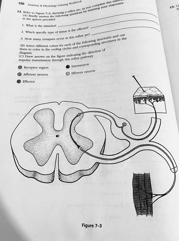
Solved 1. Which specific type of receptor is the

Information Ecology: an integrative framework for studying animal behavior: Trends in Ecology & Evolution
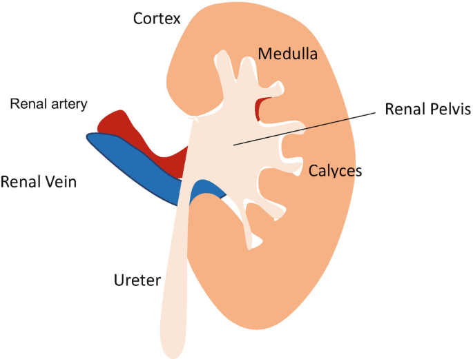
Normal Physiology of Renal System

Epigenetics - Wikipedia

Hypocretins (orexins): The ultimate translational neuropeptides - Jacobson - 2022 - Journal of Internal Medicine - Wiley Online Library

Role of Histamine and Its Receptors in Cerebral Ischemia

Dynamic θ Frequency Coordination within and between the Prefrontal Cortex-Hippocampus Circuit during Learning of a Spatial Avoidance Task

Using cognitive psychology to understand GPT-3

Breastfeeding: crucially important, but increasingly challenged in a market-driven world - The Lancet

Global investments in pandemic preparedness and COVID-19: development assistance and domestic spending on health between 1990 and 2026 - The Lancet Global Health
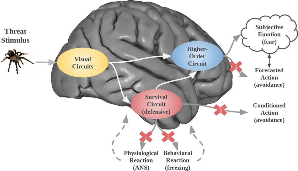
Putting the “mental” back in “mental disorders”: a perspective from research on fear and anxiety
de
por adulto (o preço varia de acordo com o tamanho do grupo)


